lv free wall rupture type | left ventricular free wall rupture lv free wall rupture type The left ventricular free-wall rupture is a serious and often lethal complication following an ST elevation myocardial infarction. However, very rarely this rupture can be . Sludinājumi. Vieglie auto - Fiat - 500, Cenas, tirdzniecība, Foto, Attēli. Visi sludinājumi
0 · ruptured left ventricular
1 · left ventricular wall rupture surgery
2 · left ventricular rupture statistics
3 · left ventricular rupture rv
4 · left ventricular rupture ncbi
5 · left ventricular free wall rupture treatment
6 · left ventricular free wall rupture
7 · free ventricular wall rupture
Finish Line | Las Vegas NV - Facebook
ruptured left ventricular
diy comment fairw un gâteau chanel
In this paper, we provide an update on the clinical, electrocardiographic, echocardiographic, and angiographic features of these patients, identifying the different forms .Between June 1993 and May 2006, 32 patients with an average age of 73 years (range, from 55 to 96 years) were surgically treated for LV free wall rupture. Sutureless technique (gluing . Becker and colleagues identified 3 morphological types of FWR. Type I rupture is characterized as an abrupt, slit-like myocardial tear and corresponds to the acute phase of . The left ventricular free-wall rupture is a serious and often lethal complication following an ST elevation myocardial infarction. However, very rarely this rupture can be .
Surgical experience for left ventricular free wall rupture after myocardial infarction has been reported, which was performed on heart beating using direct closure, large patch .
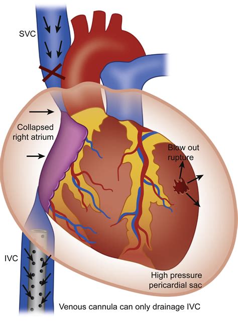
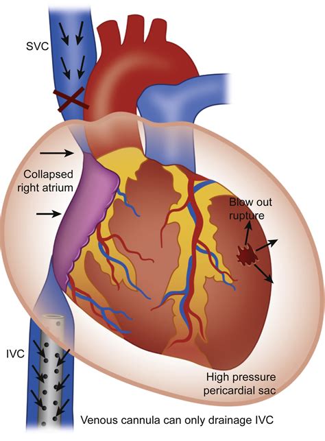
Left ventricular free wall rupture (LVFWR) is a rare complication of acute myocardial infarction (AMI), occurring in approximately 2% of cases 1 but even less frequently when primary . Patients with an oozing type of rupture located on lateral or anterolateral wall can be treated with sutureless techniques often without cardiopulmonary bypass. Patients with a blow . LV-free wall rupture is managed by resectioning the infarcted area and closure of the region with polytetrafluoroethylene or polyester patches or biological glues. Surgical repair is recommended for pseudoaneurysms, even if asymptomatic, as they carry a high risk of rupture.Left ventricular free-wall rupture (LVFWR) is an uncommon but serious mechanical complication of acute myocardial infarction. Surgical repair, though challenging, is the only definitive treatment. Given the rarity of this condition, however, results after surgery are still not well established.
Left ventricular free-wall rupture (LVFWR) may represent a dramatic and life-threatening event, occurring in up to 2% of patients with acute myocardial infarction (AMI). 1, 2 The clinical presentation varies from a catastrophic blowout type characterized by cardiogenic shock and eventually cardiac arrest, to the oozing type with haemodynamic .
In this paper, we provide an update on the clinical, electrocardiographic, echocardiographic, and angiographic features of these patients, identifying the different forms in which free wall rupture presents.Between June 1993 and May 2006, 32 patients with an average age of 73 years (range, from 55 to 96 years) were surgically treated for LV free wall rupture. Sutureless technique (gluing autologous patch to the tear) was applied in all patients. Becker and colleagues identified 3 morphological types of FWR. Type I rupture is characterized as an abrupt, slit-like myocardial tear and corresponds to the acute phase of AMI (<24 hours). In the type II rupture, an area of myocardial erosion is evident, indicating a slowly progressive tear.
left ventricular wall rupture surgery
The left ventricular free-wall rupture is a serious and often lethal complication following an ST elevation myocardial infarction. However, very rarely this rupture can be contained by the pericardium, forming a pseudoaneurysm. Surgical experience for left ventricular free wall rupture after myocardial infarction has been reported, which was performed on heart beating using direct closure, large patch and glue technique to secure homeostasis.Left ventricular free wall rupture (LVFWR) is a rare complication of acute myocardial infarction (AMI), occurring in approximately 2% of cases 1 but even less frequently when primary percutaneous intervention can be performed. 2 This complication is often fatal. Patients with an oozing type of rupture located on lateral or anterolateral wall can be treated with sutureless techniques often without cardiopulmonary bypass. Patients with a blow out type of rupture or actively squirting lesions would require a sutured approach.
LV-free wall rupture is managed by resectioning the infarcted area and closure of the region with polytetrafluoroethylene or polyester patches or biological glues. Surgical repair is recommended for pseudoaneurysms, even if asymptomatic, as they carry a high risk of rupture.
Left ventricular free-wall rupture (LVFWR) is an uncommon but serious mechanical complication of acute myocardial infarction. Surgical repair, though challenging, is the only definitive treatment. Given the rarity of this condition, however, results after surgery are still not well established. Left ventricular free-wall rupture (LVFWR) may represent a dramatic and life-threatening event, occurring in up to 2% of patients with acute myocardial infarction (AMI). 1, 2 The clinical presentation varies from a catastrophic blowout type characterized by cardiogenic shock and eventually cardiac arrest, to the oozing type with haemodynamic .
In this paper, we provide an update on the clinical, electrocardiographic, echocardiographic, and angiographic features of these patients, identifying the different forms in which free wall rupture presents.Between June 1993 and May 2006, 32 patients with an average age of 73 years (range, from 55 to 96 years) were surgically treated for LV free wall rupture. Sutureless technique (gluing autologous patch to the tear) was applied in all patients. Becker and colleagues identified 3 morphological types of FWR. Type I rupture is characterized as an abrupt, slit-like myocardial tear and corresponds to the acute phase of AMI (<24 hours). In the type II rupture, an area of myocardial erosion is evident, indicating a slowly progressive tear. The left ventricular free-wall rupture is a serious and often lethal complication following an ST elevation myocardial infarction. However, very rarely this rupture can be contained by the pericardium, forming a pseudoaneurysm.
Surgical experience for left ventricular free wall rupture after myocardial infarction has been reported, which was performed on heart beating using direct closure, large patch and glue technique to secure homeostasis.Left ventricular free wall rupture (LVFWR) is a rare complication of acute myocardial infarction (AMI), occurring in approximately 2% of cases 1 but even less frequently when primary percutaneous intervention can be performed. 2 This complication is often fatal.


Final Source Productions, Las Vegas. 108 aprecieri. Final Source Production, is the forefront supplier of LED, audio, lighting, staging, and trussing re
lv free wall rupture type|left ventricular free wall rupture


