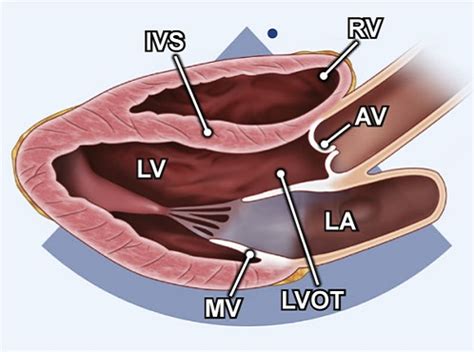
m-mode lv echocardiography plax | plax echocardiogram m-mode lv echocardiography plax PLAX view. The parasternal long axis view (PLAX) is obtained with the transducer image marker directed toward the patient’s right ear and the sound beam directed to the spine. Slight . Make a Right onto S. Las Vegas Boulevard. Our Hotel is Located on the Right just past Harrah's. Entrance is just after Denny's. Ideally located in the heart of the legendary Las Vegas Strip across from the Mirage. Only 10 minutes from McCarran International Airport.
0 · plax echocardiogram
1 · parasternal plax view echocardiogram
2 · m-mode of mv
Kad meklē lētu un pārbaudītu automašīnu, Carbuzz.lv auto placis ir īstā vieta, kur to darīt. Pie mums ir pieejamas vairāk nekā 150 dažādas automašīnas, kas atšķiras gan ar komplektāciju, gan cenu ziņā, tāpēc iespēja atrast savu nākamo sapņu auto ir ikvienam.
PLAX view. The parasternal long axis view (PLAX) is obtained with the transducer image marker directed toward the patient’s right ear and the sound beam directed to the spine. Slight .

It is used to guide M-Mode echocardiography for left ventricular measurements. Initially the parasternal long axis view is obtained. When satisfactory images are available after .
How to measure the Ejection Fraction of left ventricle using m-mode or motion mode on parasternal long axis view.
THE AMERICAN SOCIETY OF ECHOCARDIOGRAPHY RECOMMENDATIONS FOR CARDIAC CHAMBER QUANTIFICATION IN ADULTS: A QUICK REFERENCE GUIDE FROM THE ASE .
VI. M-Mode Measurements This section provides guidance on selected M-mode measurements. VII. Color Doppler Imaging This section defines the basic imaging windows, display, and mea .
Assessment of LV function with M-mode or 2-dimensional (2-D) echocardiography (Figure 2A) can be performed in the parasternal long- and short-axis views by .Use the zoom function in the PLAX view for optimal visualization of LV outflow tract (LVOT) and the aortic valve with visualization of AV cusp insertion points (annulus). Both MV leaflets and .The M-mode from the LV at the mitral valve leaflet level may be useful to measurement: a) the diastolic inter-ventricular septum, b) the diastolic posterior wall of the LV. These may be .PLAX M-mode: MV E-Septal separation (EPSS) EPSS is defined as the minimal distance between E point (most anterior motion of the AML during diastole) and a line tangential to the .
M-mode imaging in the parasternal views will further elucidate mitral leaflet motion and define the duration of mitral-septal contact. Color Doppler and PW Doppler mapping should be integrated in the assessment of .PLAX view. The parasternal long axis view (PLAX) is obtained with the transducer image marker directed toward the patient’s right ear and the sound beam directed to the spine. Slight adjustments in angle and rotation maybe necessary to demonstrate all . It is used to guide M-Mode echocardiography for left ventricular measurements. Initially the parasternal long axis view is obtained. When satisfactory images are available after fine adjustments of the transducer position, M-Mode cursor is placed in such a .
How to measure the Ejection Fraction of left ventricle using m-mode or motion mode on parasternal long axis view.THE AMERICAN SOCIETY OF ECHOCARDIOGRAPHY RECOMMENDATIONS FOR CARDIAC CHAMBER QUANTIFICATION IN ADULTS: A QUICK REFERENCE GUIDE FROM THE ASE WORKFLOW AND LAB MANAGEMENT TASK FORCE. Accurate and reproducible assessment of cardiac chamber size and function is essential for clinical care. A standardized methodology .VI. M-Mode Measurements This section provides guidance on selected M-mode measurements. VII. Color Doppler Imaging This section defines the basic imaging windows, display, and mea-surements for color Doppler imaging (CDI) to be integrated into the comprehensive transthoracic examination. Similarly, display of colorAssessment of LV function with M-mode or 2-dimensional (2-D) echocardiography (Figure 2A) can be performed in the parasternal long- and short-axis views by placing the calipers perpendicular to the ventricular long axis. Change in LV cavity dimensions during systole can be used to calculate LV fractional shortening and ejection fraction.
Use the zoom function in the PLAX view for optimal visualization of LV outflow tract (LVOT) and the aortic valve with visualization of AV cusp insertion points (annulus). Both MV leaflets and 2 of the 3 aortic leaflets should be visible in good quality.
hermes scarf alice shirley
PLAX M-mode: MV E-Septal separation (EPSS) EPSS is defined as the minimal distance between E point (most anterior motion of the AML during diastole) and a line tangential to the most posterior excursion of the IVS within the same cardiac cycle
M-mode imaging in the parasternal views will further elucidate mitral leaflet motion and define the duration of mitral-septal contact. Color Doppler and PW Doppler mapping should be integrated in the assessment of obstruction and, when present, determine the . Rapidly moving structures such as the aortic valve and mitral valve, and endocardium have characteristic movements in M-mode. M-mode also has a great spatial resolution, which is useful for measuring ventricular dimensions in systole and diastole.
plax echocardiogram
PLAX view. The parasternal long axis view (PLAX) is obtained with the transducer image marker directed toward the patient’s right ear and the sound beam directed to the spine. Slight adjustments in angle and rotation maybe necessary to demonstrate all . It is used to guide M-Mode echocardiography for left ventricular measurements. Initially the parasternal long axis view is obtained. When satisfactory images are available after fine adjustments of the transducer position, M-Mode cursor is placed in such a .How to measure the Ejection Fraction of left ventricle using m-mode or motion mode on parasternal long axis view.
THE AMERICAN SOCIETY OF ECHOCARDIOGRAPHY RECOMMENDATIONS FOR CARDIAC CHAMBER QUANTIFICATION IN ADULTS: A QUICK REFERENCE GUIDE FROM THE ASE WORKFLOW AND LAB MANAGEMENT TASK FORCE. Accurate and reproducible assessment of cardiac chamber size and function is essential for clinical care. A standardized methodology .VI. M-Mode Measurements This section provides guidance on selected M-mode measurements. VII. Color Doppler Imaging This section defines the basic imaging windows, display, and mea-surements for color Doppler imaging (CDI) to be integrated into the comprehensive transthoracic examination. Similarly, display of colorAssessment of LV function with M-mode or 2-dimensional (2-D) echocardiography (Figure 2A) can be performed in the parasternal long- and short-axis views by placing the calipers perpendicular to the ventricular long axis. Change in LV cavity dimensions during systole can be used to calculate LV fractional shortening and ejection fraction.Use the zoom function in the PLAX view for optimal visualization of LV outflow tract (LVOT) and the aortic valve with visualization of AV cusp insertion points (annulus). Both MV leaflets and 2 of the 3 aortic leaflets should be visible in good quality.
PLAX M-mode: MV E-Septal separation (EPSS) EPSS is defined as the minimal distance between E point (most anterior motion of the AML during diastole) and a line tangential to the most posterior excursion of the IVS within the same cardiac cycle M-mode imaging in the parasternal views will further elucidate mitral leaflet motion and define the duration of mitral-septal contact. Color Doppler and PW Doppler mapping should be integrated in the assessment of obstruction and, when present, determine the .
parasternal plax view echocardiogram
m-mode of mv
An E-Book of Cassius Dio's 'Roman History, Vol. V' The Project Gutenberg EBook of Dio's Rome, Volume V., Books 61-76 (A.D. 54-211), by Cassius Dio This eBook is for the use of anyone anywhere at no cost and with almost no restrictions whatsoever. . 29 . Nero continued to commit many ridiculous acts, among which may be cited his .
m-mode lv echocardiography plax|plax echocardiogram


